

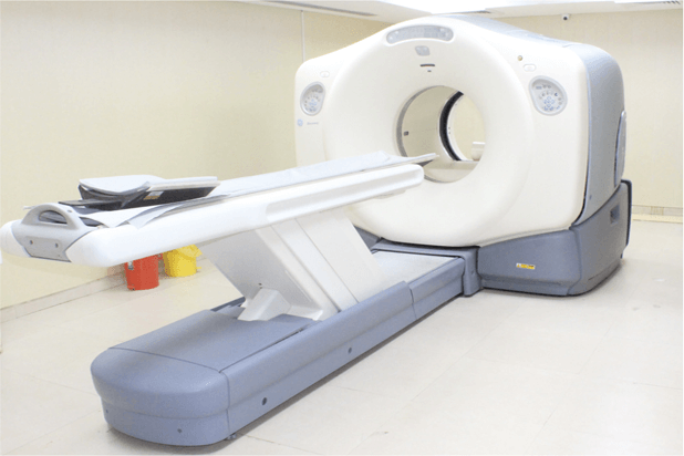
PET-CT is a combination of cross-sectional anatomic information provided by CT and metabolic information provided by positron emission tomography (PET). The basics of PET imaging is that the technique detects pairs of gamma rays emitted indirectly by a positron-emitting radionuclide (also called radiopharmaceuticals, radionuclides, or radiotracer).
The tracer Is injected into a vein on a biologically active molecule, usually a sugar that is used for cellular energy. PET systems have sensitive detector panels to capture gamma ray emissions from inside the body and use software to plot to triangulate the source of the emissions, creating 3-D computed tomography images of the tracer concentrations within the body
PET is a primary imaging modality for the detection and tracking of cancer. Nuclear imaging (PET and SPECT) are also considered a standard of care for myocardial perfusion exams to see if there is a lack of blood flow to the heart muscle due to a heart attack. PET scans can also help detect and track neurological disorders in the brain, such as Alzheimer’s disease, and the evaluation of strokes.
PET-CT has revolutionized medical diagnosis in many fields, by adding precision of anatomic localization to functional imaging, which was previously lacking from pure PET imaging. For example, many diagnostic imaging procedures in oncology, surgical planning, radiation therapy and cancer staging have been changing rapidly under the influence of PET-CT availability.
Read More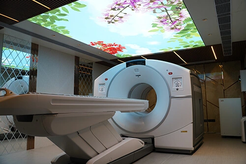
DOTA PET CT Scan (Gallium-68 DOTATATE Scan) is a sophisticated diagnostic tool for Cancer diagnosis. In this scan, a radioactive substance is injected intravenously called a tracer (Ga-68 dotatate) to detect minute damages very early on in cancer development.
DOTA PET CT Scan also aids precise cancer treatment planning and allows successful treatment response.
This is a radioactive scan that attaches itself to the tumor receptors to highlight neuroendocrine tumors.
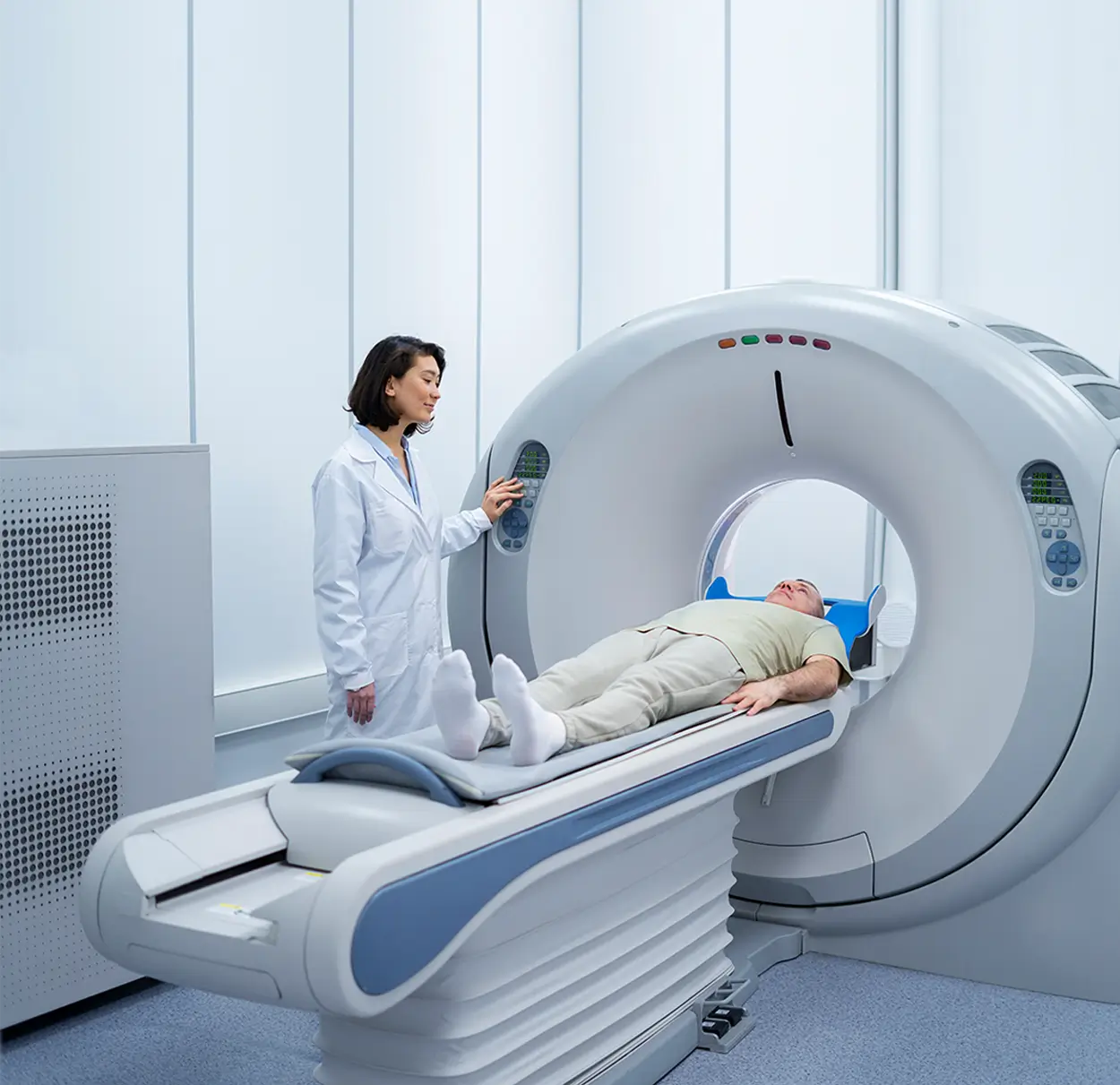
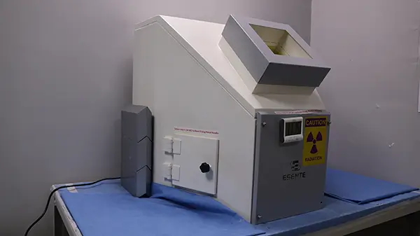
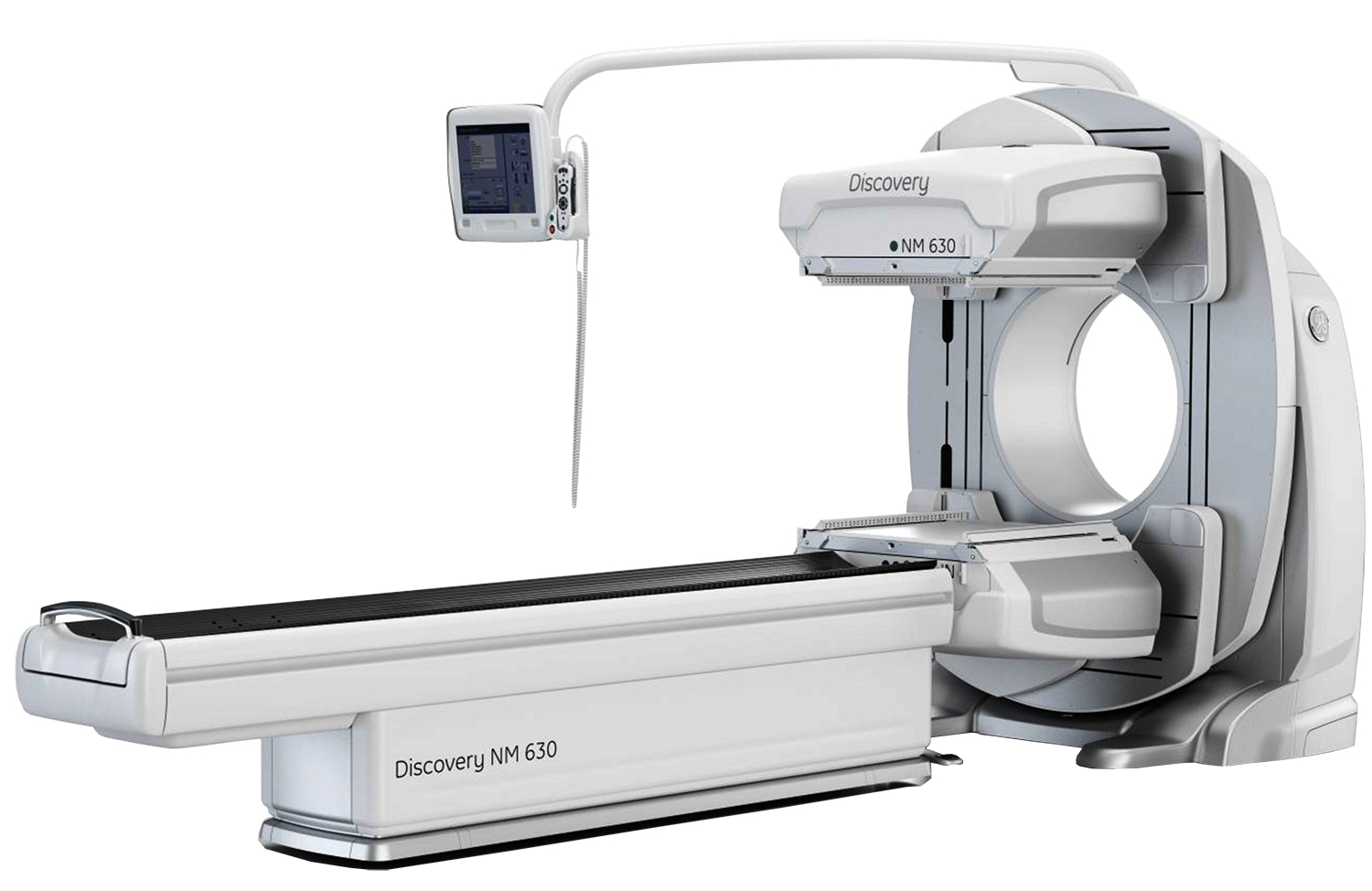
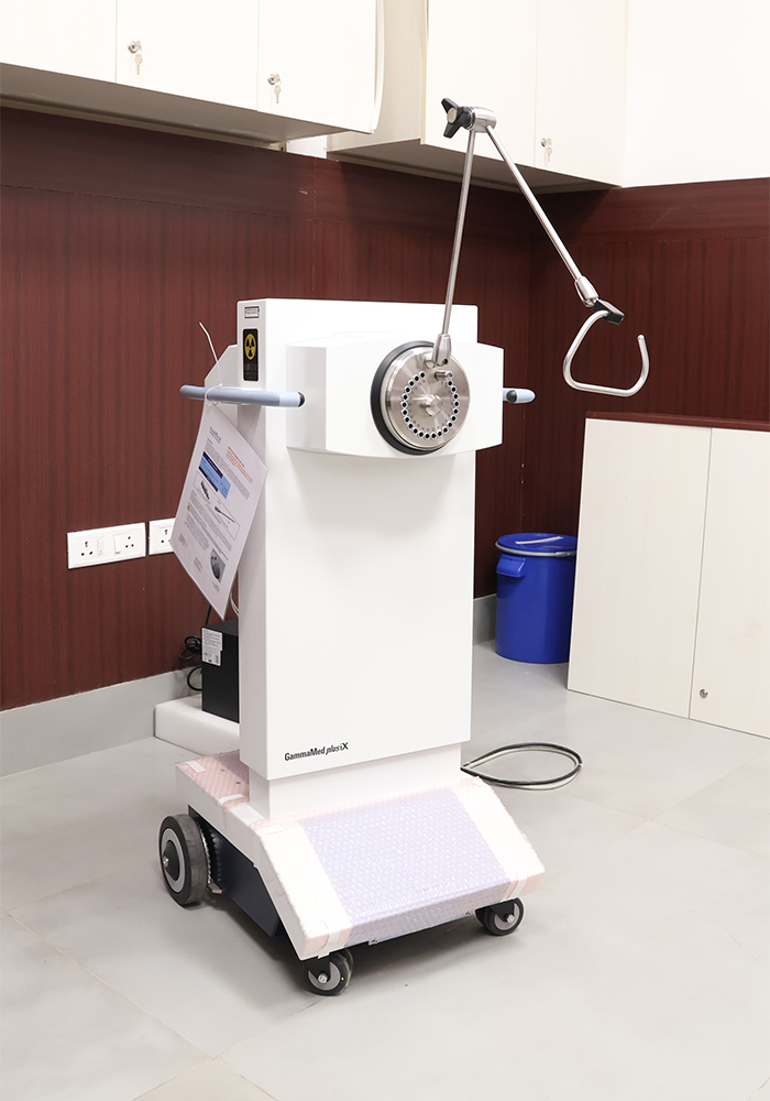
Gamma Scan analyzes the body's organs, tissue and bones through Nuclear imaging tests that use radioactive substances and a special camera to create 3D pictures. The Gamma Scan produces images that show how the organs are functioning. It also gives a clearer picture of how well blood is flowing to the heart, the areas of the brain that are active and parts of the bone that are affected by Cancer.
Read More